

NSUN5抗体NSUN5兔多抗抗体Williams-Beur
en syndrome critical region protein 20 copy A antibody抗体NSUN5 Antibody抗体- ¥440 - 1320
- CUSABIO

- CN
- CSB-PA839386LA01HU
- 2025年07月14日
- ELISA, WB; Recommended dilution: WB:1:500-1:5000
- Rabbit
- Human

企业认证
相关产品推荐更多 >

HSPE1抗体HSPE1一抗抗体Hspe1 antibody抗体HSPE1 Antibody抗体
¥880
SOX8抗体SOX8兔多抗抗体Transcription factor SOX8 antibody抗体SOX8 Antibody, Biotin conjugated抗体
¥880
TMEM243抗体TMEM243兔多抗抗体MM-TRAG抗体TMEM243 Antibody抗体
¥440MYC抗体MYC一抗抗体zc-myc antibody抗体MYC Antibody抗体
¥880
TRIM21抗体TRIM21兔多抗抗体Tripartite motif-containing protein 21 antibody抗体TRIM21 Antibody, HRP conjugated抗体
¥880
万千商家帮你免费找货
0 人在求购买到急需产品
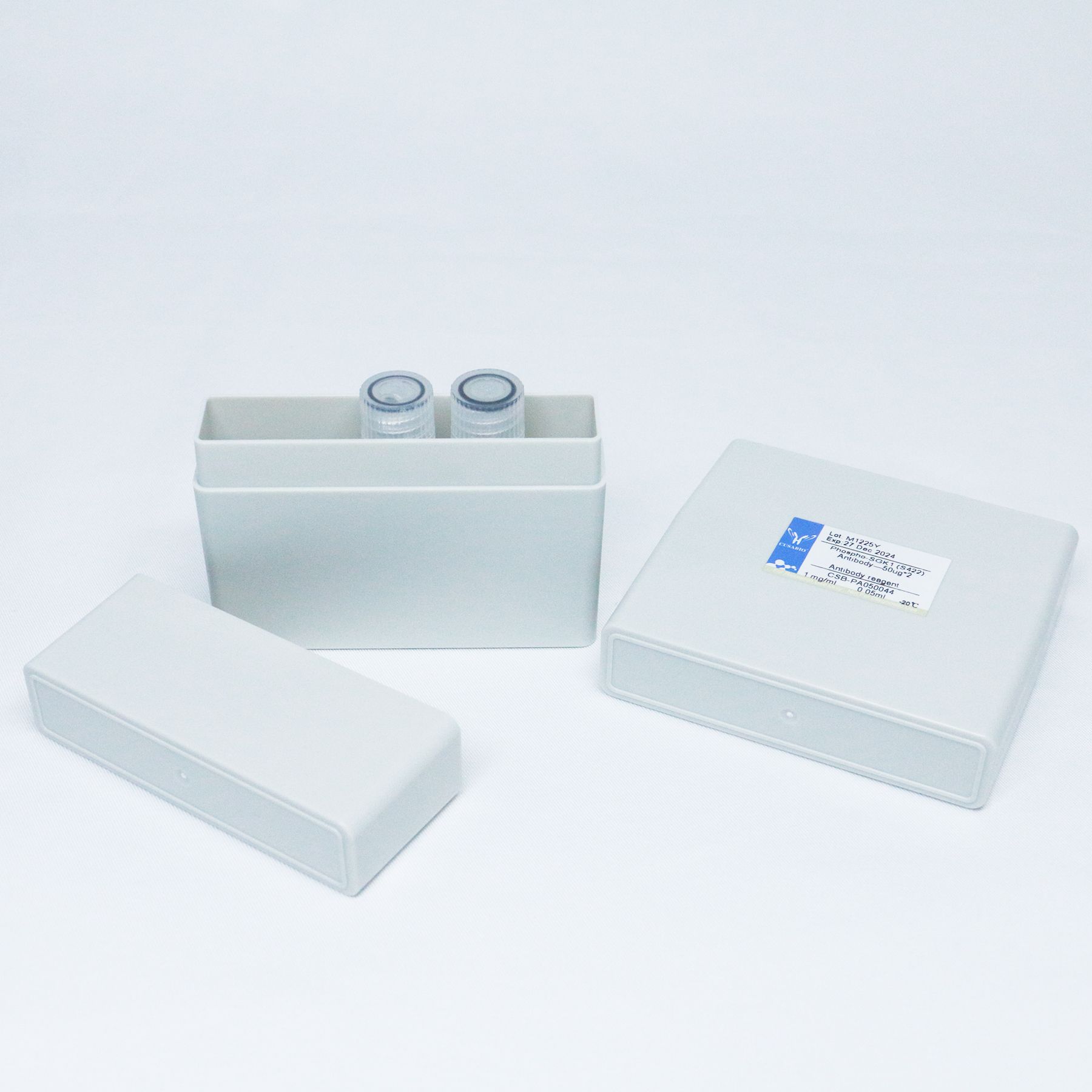
- 详细信息
- 文献和实验
- 技术资料
- 免疫原:
Recombinant Human Probable 28S rRNA (cytosine-C(5))-methyltransferase protein (362-466AA)
- 亚型:
IgG
- 形态:
Liquid
- 保存条件:
Upon receipt, store at -20℃ or -80℃. Avoid repeated freeze.
- 克隆性:
Polyclonal
- 标记物:
Non-conjugated
- 适应物种:
Human
- 保质期:
6个月
- 抗原来源:
Homo sapiens (Human)
- 目录编号:
Q96P11
- 级别:
优
- 库存:
200
- 供应商:
武汉华美生物工程有限公司
- 宿主:
Rabbit
- 应用范围:
ELISA, WB; Recommended dilution: WB:1:500-1:5000
- 浓度:
>95%,Protein G purified
- 靶点:
NSUN5
- 抗体英文名:
NSUN5 Antibody
- 抗体名:
Williams-Beuren syndrome critical region protein 20 copy A antibody
- 规格:
100μg/50μg/20μg
| 规格: | 100μg | 产品价格: | ¥1320.0 |
|---|---|---|---|
| 规格: | 50μg | 产品价格: | ¥880.0 |
| 规格: | 20μg | 产品价格: | ¥440.0 |
保存缓冲液
Preservative: 0.03% Proclin 300Constituents: 50% Glycerol, 0.01M PBS, pH 7.4
功能
S-adenosyl-L-methionine-dependent methyltransferase that specifically methylates the C(5) position of cytosine 3782 in 28S rRNA.风险提示:丁香通仅作为第三方平台,为商家信息发布提供平台空间。用户咨询产品时请注意保护个人信息及财产安全,合理判断,谨慎选购商品,商家和用户对交易行为负责。对于医疗器械类产品,请先查证核实企业经营资质和医疗器械产品注册证情况。
 文献和实验
文献和实验of many disease states, including metabolic syndrome, cardiovascular and Alzheimer's disease. Paraoxonase-1 also plays a critical role in the metabolism and detoxification of insecticides and pesticides (1). The aim of this study was to express a biologically
Mapping Networks of Protein‐Mediated Physical Interactions Between Chromatin Elements
. However, these methods do not specifically identify the protein component(s) that might mediate such interactions. This unit provides a detailed protocol for Combined 3C?ChIP?Cloning (6C) technology, which combines multiple techniques to simultaneously identify physical
Comparative Protein Structure Modeling Using MODELLER
with its model (green). (B ) Distortions and shifts in correctly aligned regions. A region in the crystal structure of mouse cellular retinoic acid binding protein I (red) is compared with its model (green) and with the template fatty acid binding protein (blue
 技术资料
技术资料暂无技术资料 索取技术资料

