大家都在搜

手机验证

询价列表

暂时没有已询价产品
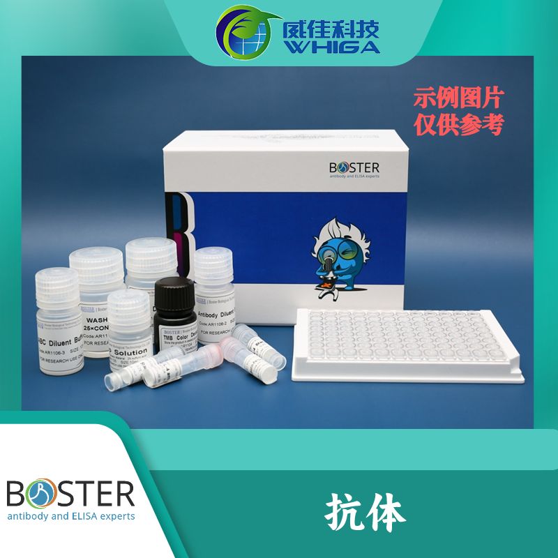
Anti-CYLD Antib
ody(PB0572-150ul)

企业认证
大量
广州威佳科技有限公司
150ul
| 货号 | PB0572 |
|---|---|
| 产品名称 | Anti-CYLD Antibody |
| 基因名 | CYLD |
| 抗体来源 | Rabbit |
| 克隆 | Polyclonal |
| 抗体亚型 | Rabbit IgG |
| 分子量 | 107KD |
| 免疫原 | E.coli-derived human CYLD recombinant protein (Position: D618-K956). Human CYLD shares 97.6% and 97.9% amino acid (aa) sequence identity with mouse and rat CYLD, respectively. |
| 内容 | 500 ug/ml antibody with PBS ,0.02% NaN3 , 1 mg BSA and 50% glycerol. |
| 纯化方式 | Immunogen affinity purified. |
| 浓度 | 500 ug/ml |
| 产品形态 | Liquid |
| 保存条件 | 12 months from date of receipt,-20℃ as supplied. 6 months 2 to 8℃ after reconstitution. Avoid repeated freezing and thawing. |
| 背景资料 | CYLD is localized on the long arm of chromosome 16. This gene encodes a cytoplasmic protein with three cytoskeletal-associated protein-glycine-conserved (CAP-GLY) domains that functions as a deubiquitinating enzyme. Mutations in this gene have been associated with cylindromatosis, multiple familial trichoepithelioma, and Brooke-Spiegler syndrome. Alternate transcriptional splice variants, encoding different isoforms, have been characterized. |
| 研究类别 | 1. Hayashi M, Jono H, Shinriki S, Nakamura T, Guo J, Sueta A, Tomiguchi M, Fujiwara S, Yamamoto-Ibusuki M, Murakami K, Yamashita S, Yamamoto Y, Li JD, Iwase H, Ando Y. “Clinical significance of CYLD downregulation in breast cancer”. Breast Cancer Res Treat, 2014 Feb.2. Vanecek T, Halbhuber Z, Kacerovska D, Martinek P, Sedivcova M, Carr RA, Slouka D, Michal M, Kazakov DV. “Large germline deletions of the CYLD gene in patients with Brooke-Spiegler syndrome and multiple familial trichoepithelioma”. Am J Dermatopathol, 2014 Nov. |
| Uniprot ID | CYLD: Q9NQC7 |
| 推荐配套的二抗和检测试剂 | Boster recommends Enhanced Chemiluminescent Kit with anti-Rabbit IgG (EK1002) for Western blot, and HRP Conjugated anti-Rabbit IgG Super Vision Assay Kit (SV0002-1) for IHC(P). *Blocking peptide 可以联系我们购买。 |
为了提供优质的抗体,博士德对每一批抗体都用没有转染过的细胞系和体细胞组织检测,以保证博士德出品的抗体有足够的亲和性足以和对应蛋白天然的表达含量起反应。
| 应用 | 稀释度* |
|---|---|
| Western blot(WB): | 1:500-2000 |
*稀释度需要用户自己调试,此处数据仅供参考。
**博士德提供高敏感的二抗和检测试剂盒。详情见相关产品推荐。
风险提示:丁香通仅作为第三方平台,为商家信息发布提供平台空间。用户咨询产品时请注意保护个人信息及财产安全,合理判断,谨慎选购商品,商家和用户对交易行为负责。对于医疗器械类产品,请先查证核实企业经营资质和医疗器械产品注册证情况。


