


- ¥1280
- 康瑞纳
- 进口/国产
- A1522
- 2025年07月16日

企业认证
相关产品推荐更多 >


万千商家帮你免费找货
0 人在求购买到急需产品
- 详细信息
- 文献和实验
- 技术资料
- 保存条件:
RT/2-8℃/-20℃
- 保质期:
3年
- 英文名:
CFDA
- 库存:
999
- 供应商:
北京康瑞纳生物科技有限公司
- CAS号:
150347-59-4
- 规格:
25mg
CFDA, 5(6)-羧基二乙酸荧光素琥珀酰亚胺酯 150347-59-4 CFDA, 5(6)-羧基二乙酸荧光素琥珀酰亚胺酯 150347-59-4
产品名称:CFDA, 5(6)-羧基二乙酸荧光素琥珀酰亚胺酯 150347-59-4
产品编号:A1522中文别称 5(6)-羧基二乙酸荧光素琥珀酰亚胺酯;CFDA, 5(6)-羧基二乙酸荧光素琥珀酰亚胺酯
英文别称 CFDA, SE [5-(and 6)-Carboxyfluorescein diacetate, succinimidyl ester] Mixed isomers CFSE
CAS 150347-59-4
分子式 C29H19NO11
分子量 557.46
储存条件 -20°C干燥避光保存 , 保质期 1年
纯度 ≥95%(HPLC)
性状:白色或浅黄色粉末。
包装 瓶
规格 25mg
产品描述:
CFDA, SE是一种可对活细胞进行荧光标记的细胞染色试剂,不仅用于细胞增殖的体外实验,也可用于追踪细胞在体内的分裂增殖过程。它穿透细胞膜进入细胞后被细胞内的酯酶催化分解成CFSE,CFSE可以偶发性地并不可逆地与细胞内的氨基结合偶联到细胞蛋白质上。在细胞分裂增殖过程中,CFSE 标记荧光可平均分配至两个子代细胞中,因此其荧光强度是亲代细胞的一半,用流式细胞仪或荧光显微镜在490 nm 激发光处可对其进行分析。CFDA, SE标记后的细胞用于体内观察可以长达数周。因此,CFDA, SE常被用来做活细胞检测实验和用荧光电镜观察细胞长期活动的实验。CFDA, SE标记的细胞的激发的发射波长分别为500nm和520nm。
外观:白色或浅黄色粉末。溶剂DMSO ,Ex (nm) 494 Em (nm) 521
染色步骤(仅供参考):
(1) 用 DMSO 制备 1mM 的 CFDA, SE 溶液。用 PBS 或适当的缓冲液将其稀释成10~50μM的CFDA,SE 溶液。
(2) 将 1/10 细胞培养基体积的 CFDA, SE 溶液加入到细胞培养基中。
(3) 在 37℃培养细胞 15~30 分钟。
(4) 用 PBS 或适当的缓冲液洗涤细胞两次。
(5) 用 490nm 激发波长、530nm 发射波长的滤光片的荧光显微镜观察细胞。
Product Description:
CFDA, SE is a cell staining reagent that can fluorescently label living cells. It is not only used for in vitro experiments on cell proliferation, but also for tracking the process of cell division and proliferation in vivo. After penetrating the cell membrane and entering the cell, it is catalyzed by esterases inside the cell to decompose into CFSE, which can occasionally and irreversibly bind to intracellular amino groups and couple to cell proteins. During the process of cell division and proliferation, CFSE labeled fluorescence can be evenly distributed between the two daughter cells, so its fluorescence intensity is half that of the parent cells. It can be analyzed by flow cytometry or fluorescence microscopy at 490 nm excitation light. Cells labeled with CFDA and SE can be obs.erved in vivo for several weeks. Therefore, CFDA and SE are often used for live cell detection experiments and for ob.serving long-term cell activity using fluorescence electron microscopy. The excitation emission wavelengths of CFDA and SE labeled cells are 500nm and 520nm, respectively.
Appearance: White or light yellow powder. Solvent DMSO, Ex (nm) 494 Em (nm) 521
Dyeing steps (for reference only):
(1) Prepare 1mM CFDA, SE solution using DMSO. Dilute it to 10-50 with PBS or appropriate buffer solution μ CFDA and SE solutions of M.
(2) Add 1/10 volume of CFDA and SE solution to the cell culture medium.
(3) Cultivate cells at 37 ℃ for 15-30 minutes.
(4) Wash the cells twice with PBS or appropriate buffer.
(5) Obs.erve cells under a fluorescence microscope using a filter with an excitation wavelength of 490nm and an emission wavelength of 530nm.
实验参考图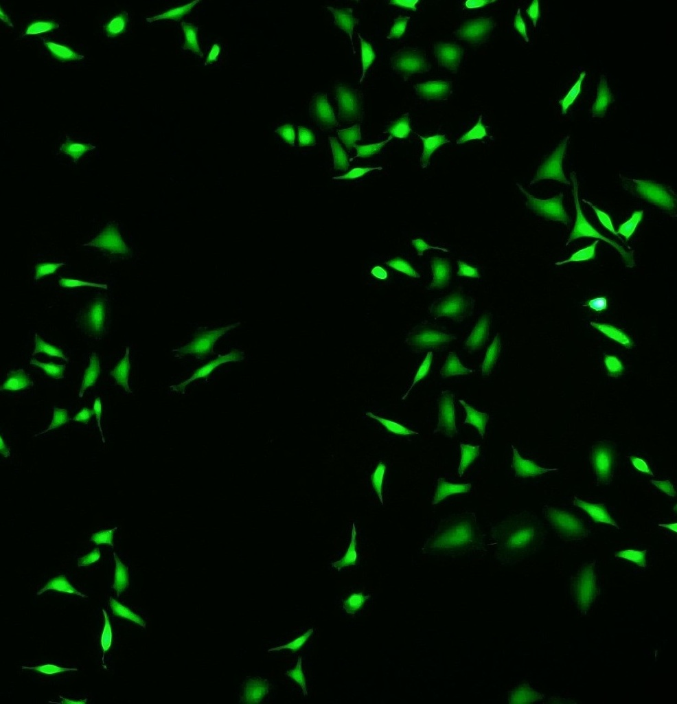
自检 20μM CFDA, SE工作液;37℃,15min;PBS 洗涤细胞两次。ExEm=490nm530nm观察细胞。
★订购流程
定购方式:
1、确认产品后可直接和我们在线联系确认您购买的产品。
2、可以把您需要购买的产品确认好以邮件的形式发给我们,这样您会在第一时间安排工作人员与您取得联系:给您回复确认的邮件,或者是电话。
邮寄产品:
现货,一般当天定购,24小时内由仓库统一出货
定制期货,出货前由专人通知,仓库备货,依其产品性能,路途等选择进行包装邮寄客户如有特别要求,可与销售人员提前沟通
质量保证与免责声明:
1、我们保证向用户提供完全合格的产品。如您认为产品确实存在质量问题,请在收到产品7天内提出,否则将不予受理或赔偿。同时产品赔偿只限于产品价值本身,不涉及其他任何损失。我们承诺随时为您提供专业的技术指导。
2、如发现产品有任何缺漏或破损,请在收到产品后2个工作日内通知我们。
3、产品使用方法: 详询客服


更多产品请详询客服
风险提示:丁香通仅作为第三方平台,为商家信息发布提供平台空间。用户咨询产品时请注意保护个人信息及财产安全,合理判断,谨慎选购商品,商家和用户对交易行为负责。对于医疗器械类产品,请先查证核实企业经营资质和医疗器械产品注册证情况。
 文献和实验
文献和实验Ren L, Liu J, Liu C, Yang T, Wu X, Zhang X, Yang L, Xia J, Li W. AIn Vitro Cell(RAW264.7 cells;100 ng/mL LPS,24 h at 37 °C) RAW264.7 cells were seeded in confocal dishes at a density of 1 × 10⁵ cells per dish. After the culture of 24 h, cells were treated with 100 ng/mL LPS in different media, including a control medium, PACDFe extract, and PACDFe hydrogel extract supplemented with 1 μM LL-37 for another 24 h at 1837 °C. 更多文献信息请详询客服
为150347-59-4。CFDA SE可以通透细胞膜 ,进入细胞后可以被细胞内的酯酶(esterase)催化分解成CFSE,CFSE可以偶发性地(spontaneously)并不可逆地和细胞内蛋白的Lysine残基或其它氨基发生结合反应,并标记这些蛋白。在加入荧光探针CFDA SE后大约24小时,即可充分标记细胞。被CFDA SE标记的非分裂细胞的荧光非常稳定,稳定标记的时间可达数个月。CFDA SE标记细胞的荧光非常均一,比以前使用的其它细胞示踪荧光探针例如PKH26的荧光更加均一,并且分裂后的子代
% 为抑制细胞毒细胞。经过适当校正,可见有两群阳性双标记细胞出现( B 图)。高强度部分为真正的 CD 8 、 CD 11 双标记阳性细胞;低强度部分属于 CD 8 绿色阳性细胞( NK 细胞)。此时 “2” 窗中细胞份额减为 7.59% 。需要指出的是校正过度则会使各区的阳性细胞都减少(图 C )。 图 10-13 双染色二维点图上游标的设置 1 区:红色荧光阳性; 2 区 :红、绿荧光均为阳性; 3 区:阴性细胞: 4 区:绿色荧光阳性


