大家都在搜

手机验证

询价列表

暂时没有已询价产品
Rabbit anti-RRM
1 Recombinant Monoclonal Antibody(R152)
企业认证
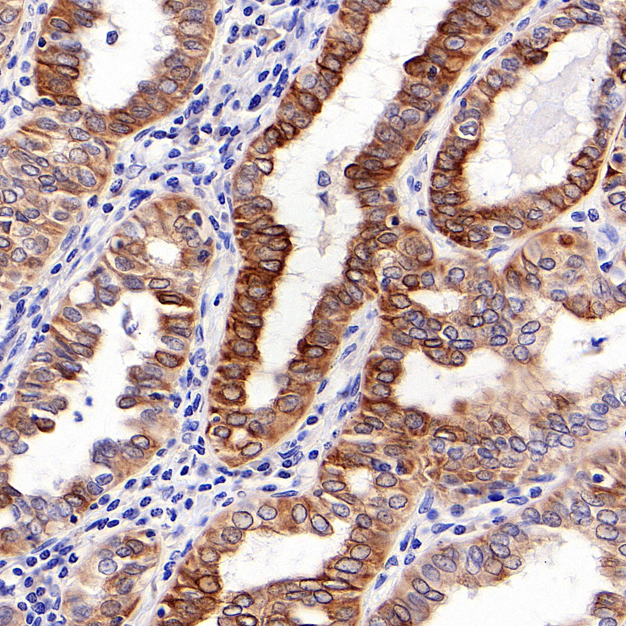
Rabbit anti-RRM1 Recombinant Monoclonal Antibody(R152)
Rabbit anti-RRM1 Recombinant Monoclonal Antibody(R152)
-
0.5 mg/mL
IHC-P: 1:100, ICC: 1:500
Rabbit
-
Store at -20℃ for one year.
-
-
-
现货
爱必信(上海)生物科技有限公司
-
-
Store at -20℃ for one year.
液态
-
-
100ul
| 概述 | |
| 别名 | Ribonucleoside-diphosphate reductase large subunit; Ribonucleoside-diphosphate reductase subunit M1; Ribonucleotide reductase large subunit |
| 宿主 | Rabbit |
| 反应种属 | Human |
| 应用 | IHC-P: 1:100, ICC: 1:500 |
| 性能 | |
| 形式 | Liquid |
| 浓度 | 0.5 mg/mL |
| 纯化方法 | Protein A affinity column |
| 类型 | Monoclonal Antibody |
| 克隆号 | R152 |
| 储存/保存方法 | Store at -20℃ for one year. |
| 存储溶液 | PBS, 40% Glycerol, 0.05% BSA, 0.03% Proclin 300 |
| 靶标 | |
| 背景说明 | The large subunit of human ribonucleotide reductase, RRM1, is involved in the regulation of cell proliferation, cell migration, tumour and metastasis development, and the synthesis of deoxyribonucleotides for DNA synthesis. It is also a cellular target for the chemotherapeutic agent, gemcitabine. RRM1 has been studied in a large number of patients with different types of cancer, such as non-small-cell lung cancer, pancreatic cancer, breast cancer, and biliary tract cancer, to establish its prognostic or predictive value when patients were treated with gemcitabine, and mRNA expression and genetic variants as determined by genotyping have in some cases been associated with clinical outcome of patients with cancer [PMID: 21163702]. |
| 细胞定位 | Cytoplasm |
| UniProt | P23921 |


风险提示:丁香通仅作为第三方平台,为商家信息发布提供平台空间。用户咨询产品时请注意保护个人信息及财产安全,合理判断,谨慎选购商品,商家和用户对交易行为负责。对于医疗器械类产品,请先查证核实企业经营资质和医疗器械产品注册证情况。