大家都在搜

手机验证

询价列表

暂时没有已询价产品

Villin antibody


企业认证
Recombinant protein encompassing a sequence within the N-terminus region of human Villin. The exact sequence is proprietary.
IgG
Liquid
Store as concentrated solution. Centrifuge briefly prior to opening vial. For short-term storage (1-2 weeks), store at 4ºC. For long-term storage, aliquot and store at -20ºC or below. Avoid multiple freeze-thaw cycles.
Polyclonal
Unconjugated
Human, Mouse, Rat, Cat, Dog
12 months from the shipping date of the product.
Human
GTX109940
Primary Antibodies
Available
GeneTex
Rabbit
WB, ICC/IF, IHC-P, IHC-Fr
0.85 mg/ml (Please refer to the vial label for the specific concentration.)
Villin
Villin antibody
Villin 抗体
100 μl/25 μl
| 规格: | 100 μl | 产品价格: | ¥4000.0 |
|---|---|---|---|
| 规格: | 25 μl | 产品价格: | ¥1700.0 |
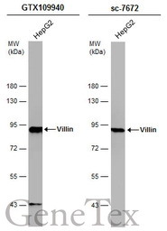
Whole cell extract (30 μg) was separated by 7.5% SDS-PAGE, and the membranes were blotted with Villin antibody (GTX109940) diluted at 1:5000 and competitor's antibody (sc-7672) diluted at 1:100. The HRP-conjugated anti-rabbit IgG antibody (GTX213110-01) was used to detect the primary antibody.
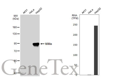
Various whole cell extracts (30 μg) were separated by 7.5% SDS-PAGE, and the membrane was blotted with Villin antibody (GTX109940) diluted at 1:2000. The HRP-conjugated anti-rabbit IgG antibody (GTX213110-01) was used to detect the primary antibody. Corresponding RNA expression data for the same cell lines are based on Human Protein Atlas program.

Villin antibody detects Villin protein at cytoplasm by immunohistochemical analysis.Sample: Paraffin-embedded rat kidney.Villin stained by Villin antibody (GTX109940) diluted at 1:500.Antigen Retrieval: Citrate buffer, pH 6.0, 15 min

Confocal immunofluorescence analysis (Olympus FV10i) of methanol-fixed HeLa, using Villin(GTX109940) antibody (Green) at 1:500 dilution. Alpha-tubulin filaments were labeled with GTX11304 (Red) at 1:2000.
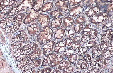
Villin antibody detects Villin protein at cytoplasm by immunohistochemical analysis.Sample: Paraffin-embedded rat colon.Villin stained by Villin antibody (GTX109940) diluted at 1:500.Antigen Retrieval: Citrate buffer, pH 6.0, 15 min
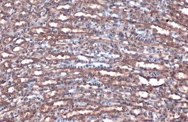
Villin antibody detects Villin protein at cytoplasm by immunohistochemical analysis.Sample: Paraffin-embedded mouse kidney.Villin stained by Villin antibody (GTX109940) diluted at 1:500.Antigen Retrieval: Citrate buffer, pH 6.0, 15 min

Villin antibody detects Villin protein at cytoplasm by immunohistochemical analysis.Sample: Paraffin-embedded mouse duodenum.Villin stained by Villin antibody (GTX109940) diluted at 1:500.Antigen Retrieval: Citrate buffer, pH 6.0, 15 min

Immunohistochemical analysis of paraffin-embedded A549 xenograft, using Villin (GTX109940) antibody at 1:500 dilution.
Antigen Retrieval: Trilogy™ (EDTA based, pH 8.0) buffer, 15min
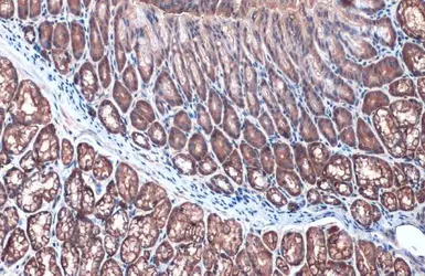
Villin antibody detects Villin protein at cytoplasm by immunohistochemical analysis.Sample: Paraffin-embedded mouse duodenum.Villin stained by Villin antibody (GTX109940) diluted at 1:500.Antigen Retrieval: Citrate buffer, pH 6.0, 15 min

Mouse tissue extract (50 μg) was separated by 7.5% SDS-PAGE, and the membrane was blotted with Villin antibody (GTX109940) diluted at 1:1000. The HRP-conjugated anti-rabbit IgG antibody (GTX213110-01) was used to detect the primary antibody.
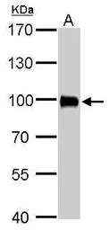
Villin antibody detects VIL1 protein by western blot analysis.
A. 50 μg rat kidney lysate/extract
7.5% SDS-PAGE
Villin antibody (GTX109940) dilution: 1:2000
The HRP-conjugated anti-rabbit IgG antibody (GTX213110-01) was used to detect the primary antibody.
风险提示:丁香通仅作为第三方平台,为商家信息发布提供平台空间。用户咨询产品时请注意保护个人信息及财产安全,合理判断,谨慎选购商品,商家和用户对交易行为负责。对于医疗器械类产品,请先查证核实企业经营资质和医疗器械产品注册证情况。