大家都在搜

手机验证

询价列表

暂时没有已询价产品

DAI/ZBP1 Antibo
dy 抗体,orb745977,Biorbyt

企业认证
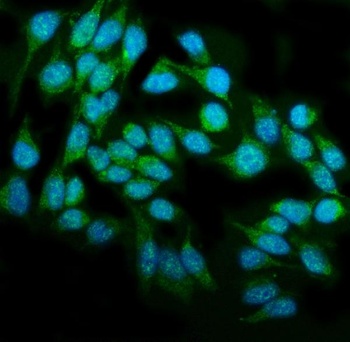
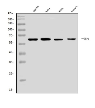
DAI/ZBP1 Antibody 抗体
DAI/ZBP1 Antibody
Adding 0.2 ml of distilled water will yield a concentration of 500 μg/ml.
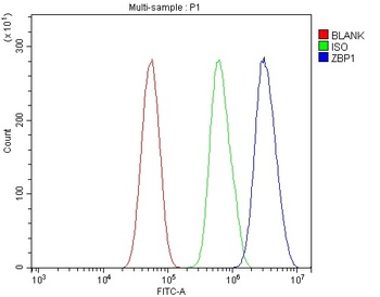
ELISA, FC, ICC, IF, WB
Rabbit
Human
6-12个月
详询
orb745977
科研
99
Biorbyt
Unconjugated
POLYCLONAL
Store at -20˚C for one year from date of receipt. After reconstitution, at 4˚C for one month. It can also be aliquotted and stored frozen at -20˚C for six months. Avoid repeated freeze-thaw cycles.
Lyophilized
Rabbit IgG
E.coli-derived human DAI/ZBP1 recombinant protein (Position: A2-E421).
10 ug/100 ug
| 规格: | 10 ug | 产品价格: | ¥3484.0 |
|---|---|---|---|
| 规格: | 100 ug | 产品价格: | ¥6760.0 |
风险提示:丁香通仅作为第三方平台,为商家信息发布提供平台空间。用户咨询产品时请注意保护个人信息及财产安全,合理判断,谨慎选购商品,商家和用户对交易行为负责。对于医疗器械类产品,请先查证核实企业经营资质和医疗器械产品注册证情况。