

- ¥1280 - 1980
- YLKBIO
- YLK-KT2010D
- 国产
- 2026年01月13日
- WB=1:100-500 ELISA=1:500-1000 IHC-P=1:100-500 IHC-F=1:100-500 ICC=1:100-500 IF=1:100-500
- Human, Mouse, Rat, Cow, Sheep, Chimpanzee
- /

企业认证
相关产品推荐更多 >


万千商家帮你免费找货
0 人在求购买到急需产品
- 详细信息
- 文献和实验
- 技术资料
- 供应商:
优利科(上海)生命科学有限公司
- 库存:
130
- 靶点:
详询
- 级别:
科研
- 目录编号:
/
- 克隆性:
多克隆
- 抗原来源:
详见说明书
- 保质期:
1年
- 抗体英文名:
Anti-Inversin/Nephrocystin 2
- 抗体名:
Inversin/Nephrocystin 2
- 标记物:
详询
- 宿主:
Human, Mouse, Rat, Cow, Sheep, Chimpanzee
- 适应物种:
/
- 免疫原:
KLH conjugated synthetic peptide derived from human Inversin/Nephrocystin 2
- 亚型:
IgG
- 形态:
Lyophilized or Liquid
- 应用范围:
WB=1:100-500 ELISA=1:500-1000 IHC-P=1:100-500 IHC-F=1:100-500 ICC=1:100-500 IF=1:100-500
- 保存条件:
-20℃
- 浓度:
1mg/ml
- 规格:
详询

| 英文名称 Anti-Inversin/Nephrocystin 2 |
| 中文名称 内脏器官发育转位相关蛋白NPH2抗体 |
| 别 名 INV; Inversion of embryo turning homolog; inversion of embryonic turning; INVS; Nephrocystin 2; Nephrocystin-2; Nephrocystin2; nephronophthisis 2 (infantile); NPH2; NPH-P2; INVS_HUMAN. |
| 浓 度 1mg/1ml |
| 规 格 0.2ml/200μg |
| 抗体来源 Rabbit |
| 克隆类型 polyclonal |
| 交叉反应 Human, Mouse, Rat, Cow, Sheep, Chimpanzee |
| 产品类型 一抗 |
| 研究领域 细胞生物 发育生物学 信号转导 干细胞 |
| 蛋白分子量 predicted molecular weight: 118kDa |
| 性 状 Lyophilized or Liquid |
| 免 疫 原 KLH conjugated synthetic peptide derived from human Inversin/Nephrocystin 2 |
| 亚 型 IgG |
纯化方法 affinity purified by Protein A 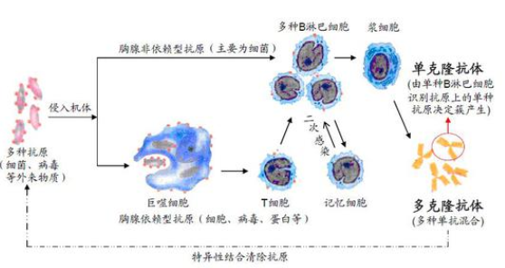 |
| 储 存 液 Preservative: 15mM Sodium Azide, Constituents: 1% BSA, 0.01M PBS, pH 7.4 |
| 产品应用 WB=1:100-500 ELISA=1:500-1000 IHC-P=1:100-500 IHC-F=1:100-500 ICC=1:100-500 IF=1:100-500 |
| (石蜡切片需做抗原修复) |
| not yet tested in other applications. |
| optimal dilutions/concentrations should be determined by the end user. |
| 保存条件 Store at -20 °C for one year. Avoid repeated freeze/thaw cycles. The lyophilized antibody is stable at room temperature for at least one month and for greater than a year when kept at -20°C. When reconstituted in sterile pH 7.4 0.01M PBS or diluent of antibody the antibody is stable for at least two weeks at 2-4 °C. |
| Important Note This product as supplied is intended for research use only, not for use in human, therapeutic or diagnostic applications. |
| 产品介绍 Nephrocystin-2 is a 1,065 amino acid protein that exists as three alternatively spliced isoforms and is essential for establishment of the left-right axis and normal renal development. Localizing to the cytoplasm, cytoskeleton, membrane and nucleus, nephrocystin-2 is expressed during presomite-stage embryos and persists in adulthood, with high levels of expression in liver and kidney. Mice expressing nephrocystin-2 mutations are primarily generated by random insertional mutagenesis and result in the reversal of left/right polarity and cyst formation in the kidneys. Furthermore, altered nephrocystin-2 function reverses nodal and lefty expression, indicating that nephrocystin-2 signaling occurs upstream of these proteins involved in the development of asymmetry. |
| Function : Required for normal renal development and establishment of left-right axis. Probably acts as a molecular switch between different Wnt signaling pathways. Inhibits the canonical Wnt pathway by targeting cytoplasmic disheveled (DVL1) for degradation by the ubiquitin-proteasome. This suggests that it is required in renal development to oppose the repression of terminal differentiation of tubular epithelial cells by Wnt signaling. Binds calmodulin via its IQ domains. Interacts with microtubules. (from SwissProt). |
| Subunit : Binds calmodulin via its IQ domains. Interacts with APC2. Interacts with alpha-, beta-, and gamma-catenin. Interacts with N-cadherin (CDH2). Interacts with microtubules (By similarity). Interacts with NPH-P1. Interacts with DVL1, PRICKLE (PRICKLE1 or PRICKLE2) and Strabismus (VANGL1 or VANGL2). Interacts with NPH-P3. Interacts with IQCB1; the interaction likely requires additional interactors. |
| Subcellular Location : Cytoplasm, cytoskeleton, spindle, membrane; Peripheral membrane protein, nucleus. Note=Associates with several components of the cytoskeleton including ciliary, random and polarized microtubules. During mitosis, it is recruited to mitotic spindle. Frequently membrane-associated, membrane localization is dependent upon cell-cell contacts and is redistributed when cell adhesion is disrupted after incubation of the cell monolayer with low-calcium/EGTA medium. |
| Tissue Specificity : Widely expressed. Strongly expressed in the primary cilia of renal tubular cells. |
| Post-translational modifications : May be ubiquitinated via its interaction with APC2 (By similarity). |
DISEASE : Defects in INVS are the cause of nephronophthisis type 2 (NPH-P2) [MIM:602088]; also known as infantile nephronophthisis. NPH-P2 is an autosomal recessive disorder resulting in end-stage renal disease. It is characterized by early onset and rapid progression. Phenotypic manifestations include enlarged kidneys, chronic tubulo-interstitial nephritis, anemia, hyperkalemic metabolic acidosis. Some patients also display situs inversus. Pathologically, it differs from later-onset nephronophthisis by the absence of medullary cysts and thickened tubular basement membranes and by the presence of cortical microcysts.  |
| Similarity : Contains 16 ANK repeats. |
| Contains 2 IQ domains. |
Database links : UniProtKB/Swiss-Prot: Q9Y283.2 |
| AGER(Advanced glycosylation end product-specific receptor) 晚期糖基化终末产物特异性受体(抗原) |
| 14-3-3 protein 14-3-3 蛋白(多肽抗原) |
| AIF (Apoptosis-Inducing Factor) 调亡诱导因子(多肽片断抗原) |
| Adenylate Kinase 1 (AK-1) 腺苷酸激酶-1 |
| AIF (Apoptosis-Inducing Factor) 调亡诱导因子(多肽片断抗原) |
| Akt/PKB 蛋白激酶B(抗原) |
| 2,4-D/KLH(2,4-Dichlorophenoxyacetic acid) 2,4-二氯苯氧乙酸偶联血蓝蛋白 |
| Akt/PKB 蛋白激酶B(抗原) |
| ALK (Anaplastic Lymphoma Kinase)(CD246Ag) 间变型淋巴瘤激酶(抗原) |
| AM Adrenomedullin 肾上腺髓质素(抗原) |
| TIMP-2(Tissue Inhibitor of Metalloproteinase-2) 金属蛋白酶组织抑制因子-2(抗原) |
| TIMP-3(Tissue Inhibitor of Metalloproteinase-3) 金属蛋白酶组织抑制因子(抗原) |
| ADM R, Adrenomedullin receptor 肾上腺髓质素受体(抗原) |
| Amylin1-37/FITC 异硫QING酸荧光素(FITC)标记糖尿病相关肽1-37 |
| VTG(vitellogenin) 青鳉鱼卵黄蛋白 |
| Apaf-1 (Apoptosis protease activating factor-1)(CT) 凋亡蛋白活性因子-1(抗原) |
风险提示:丁香通仅作为第三方平台,为商家信息发布提供平台空间。用户咨询产品时请注意保护个人信息及财产安全,合理判断,谨慎选购商品,商家和用户对交易行为负责。对于医疗器械类产品,请先查证核实企业经营资质和医疗器械产品注册证情况。
 文献和实验
文献和实验凋亡相关蛋白TFAR19 在TF21细胞凋亡中出现细胞核转位
凋亡相关蛋白TFAR19 在TF21 细胞凋亡中出现细胞核转位 陈英玉,孙荣华,韩文玲,张颖妹,宋泉声,狄春辉,马大龙 (北京大学人类疾病基因研究中心,北京 100083) [关键词] TFAR19 ; TF21 细胞;凋亡;核转位;核蛋白类 [摘 要] 目的:探讨凋亡相关分子TFAR19 在TF21 细胞凋亡过程中的表达及定位。方法
自己知识产权的人类新基因,前期的功能研究表明,它是促进细胞凋亡的增强剂.利用荧光 素(FITC)标记的TFAR19单克隆抗体为探针,对细胞凋亡过程中TFAR19蛋白的表达水平及定位研究发现,凋亡早期TFAR19表达水平增高并出 现快速核转位现象...查看原文 了解更多有用的、有意思的前沿资讯以及酷炫的实验方法,可以向大师兄申请微信好友 师兄微信号:shixiongcoming
,内含胞浆成分,如线粒体、内质网、核糖体等。 自噬溶酶体(AV2)的特征为:单层膜,胞浆成分已降解。(autophagic vacuole,AV) 2. 在荧光显微镜下采用 GFP-LC3 融合蛋白来示踪自噬形成 由于电镜耗时长,不利于监测(Monitoring)自噬形成,人们利用 LC3 在自噬形成过程中发生聚集的现象开发出了此技术。无自噬时,GFP-LC3 融合蛋白弥散在胞浆中;自噬形成时,GFP-LC3 融合蛋白转位至自噬体膜,在荧光显微镜下形成多个明亮的绿色荧光斑点,一个斑


