

- 询价
- Biotium

- 美国
- 30021
- 2026年01月17日

企业认证
相关产品推荐更多 >

万千商家帮你免费找货
0 人在求购买到急需产品
- 详细信息
- 文献和实验
- 技术资料
- 保存条件:
4°C
- 保质期:
12个月
- 英文名:
CELLBRITE GREEN CYTOPLASMIC MEMBRANE DYE 30021
- 库存:
99
- 供应商:
Biotium
- CAS号:
1
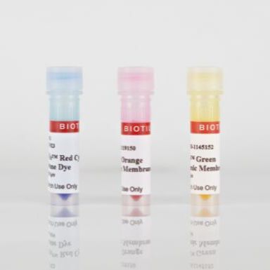
CellBrite® Cytoplasmic Membrane Labeling Kits can be used to label cell cytoplasmic membranes with fluorescent dyes, available in blue, green, orange, and red. The labeling is nontoxic and suitable for long-term tracking of cells. The dyes also can be used to stain membranes of formaldehyde-fixed cells.
Features
- Lipophilic carbocyanine dye solutions
- Low toxicity and minimal dye transfer between cells
- Useful for cell tracing, tracking, and transplantation studies
- Formaldehyde-fixable
- Stain live cells in culture medium, or stain formaldehyde-fixed cells
Kit Components
- Optimized dye stock solution
- Loading buffer (CellBrite® Blue only)
Spectral Properties
- CellBrite® Blue: Ex/Em 366/441 nm
- CellBrite® Green: Ex/Em 484/501 nm
- CellBrite® Orange: Ex/Em 549/565 nm
- CellBrite® Red: Ex/Em 644/665 nm
CellBrite® stains are based on lipophilic carbocyanine dyes: CellBrite® Blue is based on DiB, CellBrite® Green is based on Neuro-DiO, CellBrite® Orange is based on DiI, and CellBrite® Red is based on DiD. Carbocyanine dyes label cytoplasmic membrane and intracellular membrane structures efficiently and permanently. They have been used as tracers in cell fusion, cell adhesion, and cell migration applications due to their properties of low cytotoxicity and high resistance to intercellular transfer. By combining multiple CellBrite® Cytoplasmic Membrane Stains, one can label multiple cell populations with different colors for studies of cell-cell interactions.
Note: CellBrite® dyes primarily localize to cell surface membranes shortly after staining. However, in live cells the stained membranes become internalized by endocytosis, so staining becomes primarily intracellular over the course of a few hours in culture. See “Find the Right Stain for Your Application” below, and our Membrane Staining & Imaging Tech Tip to learn more.
Biotium’s CellBrite® Cytoplasmic Membrane Dyes are dye delivery solutions that can be added directly to normal culture media to uniformly label suspended or adherent cells in culture. CellBrite® dyes allow cell populations to be marked in distinctive fluorescent colors for identification after mixing. Double labeling can identify cells that have fused or formed stable clusters.
We also offer CellBrite® NIR Cytoplasmic Membrane Dyes with emission in the far-red/near-infrared region for imaging by either microscopy or near-infrared imaging devices.
Find the Right Stain for Your Application
CellBrite® dyes are non-toxic and stain cells very stably. They can be used to track cells for days to weeks. However, over time the dyes will be internalized by endocytosis, resulting in labeling of intracellular vesicles. A few hours after staining, the dyes will no longer outline the plasma membrane, but will be localized inside the cell. For long-term visualization of cell morphology in culture, our stable, non-toxic ViaFluor® SE Cell Proliferation Kits may be a more suitable alternative. ViaFluor® SE dyes covalently label cells throughout the cytoplasm and can be tracked for several days or longer by microscopy or flow cytometry. See our Tech Tip: Using ViaFluor® SE Stains for Cell Tracing and Co-Culture to learn more.
CellBrite® dyes can be used to stain live cells or formaldehyde-fixed cells. Live cells can be fixed in formaldehyde after staining as well, but the staining has poor tolerance for methanol fixation or detergent permeabilization. However, permeabilizing fixed cells before staining with CellBrite® dyes can give good results, see our Tech Tip: Combining Lipophilic Membrane Dyes with Immunofluorescence. For membrane stains that withstand fixation with either formaldehyde or methanol, as well as detergent permeabilization after staining, see our CellBrite® Fix Membrane Stains and MemBrite® Fix Cell Surface Staining Kits.
CellBrite® dyes do not stain yeast or bacteria. See our Cellular Stains Table for more information on how our dyes stain various organisms.
Product Information
| Catalog No. | Membrane Dye | Abs/Em |
|---|---|---|
| 30024 | CellBrite™ Blue | 366/441 nm |
| 30021 | CellBrite™ Green | 484/501 nm |
| 30022 | CellBrite™ Orange | 549/565 nm |
| 30023 | CellBrite™ Red | 644/665 nm |
风险提示:丁香通仅作为第三方平台,为商家信息发布提供平台空间。用户咨询产品时请注意保护个人信息及财产安全,合理判断,谨慎选购商品,商家和用户对交易行为负责。对于医疗器械类产品,请先查证核实企业经营资质和医疗器械产品注册证情况。
 文献和实验
文献和实验analysis of human lymphoblastoid cell lines (LCL) for relative quantitation of mitochondrial-localized fluorescent dye intensity. The specific dyes described include MitoTracker Green FM to assess mitochondrial content, tetramethylrhodamine ethyl ester
Synthesis and Probing of Membrane-bound Peptide Arrays
Synthesis and Probing of Membrane-bound Peptide Arrays Ronald Frank Department of Chemical Biology, GBF (German Research Center for Biotechnology), 38124 Braunschweig, Germany
Two dimensional peptide mapping
of the plate, 2.5 cm up from the bottom. For phosphorylated Lck, run pH 8.9 map at 1KV for 27 min. in the 1st dimension. 11. It is important to run marker dyes. The green dye is a 1:1 mixture of Xylene cyanol FF


