

- 询价
- Biotium

- 美国
- 70065-T
- 2026年01月23日

企业认证
相关产品推荐更多 >

万千商家帮你免费找货
0 人在求购买到急需产品
- 详细信息
- 文献和实验
- 技术资料
- 保存条件:
4°C避光
- 保质期:
12个月
- 英文名:
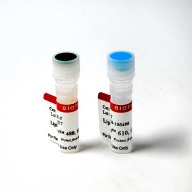
LipidSpot™ 488 Lipid Droplet Stain,1000X in DMSO Biotium 70065-T 70065-T
- 库存:
99
- 供应商:
Biotium
- CAS号:
1
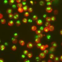
LipidSpot™ stains are fluorescent dyes that rapidly stain lipid droplets in live or fixed cells, with no wash step and minimal background.
- Rapidly and specifically stain lipid droplets
- Minimal background, no wash required
- Stain live or fixed cells, or fix and permeabilize after staining
- Suitable for staining 3D cell spheroids
- Available with green or red/far-red fluorescence
- LipidSpot™ 488 validated in super-resolution imaging by SIM
- Supplied at 1000X in DMSO
Intracellular lipid droplets are cytoplasmic organelles involved in the storage and regulation of triglycerides and cholesterol esters. LipidSpot™ dyes are fluorogenic neutral lipid stains that rapidly accumulate in lipid droplets, where they become brightly fluorescent. The dyes can be used to stain lipid droplets in both live and fixed cells, with no wash step required. Cells also can be fixed and permeabilized after staining. LipidSpot™ stains show minimal background staining of cellular membranes or other organelles, unlike traditional dyes like Nile Red.
Find the Right Stain for Your Application
LipidSpot™ 488 has excitation around 430 nm, and can be excited equally well at 405 nm or 488 nm. In cells, it stains lipid droplets with bright green fluorescence detectable in the FITC channel. LipidSpot™ 488 has been validated in super-resolution imaging by SIM (Ref. 3), and for staining of 3-D cell spheroids (Ref. 7).
LipidSpot™ 610 has excitation/emission at ~592/638 nm in cells; it is optimally detected in the Texas Red® channel, but is also bright in the Cy®3 and far-red Cy®5 channels. Therefore, we don’t recommend pairing LipidSpot™ 610 with other red or far-red probes.
In yeast, LipidSpot™ 488 stains intracellular membranes, but LipidSpot™ 610 does not. In bacteria, both LipidSpot™ dyes can stain gram-positive but not gram-negative strains. See our Cellular Stains Table for more information on how our dyes stain various organisms.
LipidSpot™ Lipid Droplet Stains
| LipidSpot™ Stain | Abs/Em | Detection channel | Catalog no. | Size (1000X in DMSO) |
|---|---|---|---|---|
| LipidSpot™ 488 | 427/585 nm (in vegetable oil or cells) |
FITC, GFP | 70065-T | 20 uL |
| 70065 | 125 uL | |||
| LipidSpot™ 610 | 610/663 nm (in vegetable oil) ~592/638 nm (in cells) |
Texas Red® or Cy®5 |
70069-T | 20 uL |
| 70069 | 125 uL |
风险提示:丁香通仅作为第三方平台,为商家信息发布提供平台空间。用户咨询产品时请注意保护个人信息及财产安全,合理判断,谨慎选购商品,商家和用户对交易行为负责。对于医疗器械类产品,请先查证核实企业经营资质和医疗器械产品注册证情况。
 文献和实验
文献和实验Vybrant® DyeCycle Green and Orange Stains
Stability Vybrant® DyeCycle™ Green stain Vybrant® DyeCycle™ Orange stain 400 μL 5 mM solution in dimethyl sulfoxide (DMSO
人 iPS 细胞体外分化为气道上皮肺类器官用于呼吸系统疾病研究应用以及iPSC操作指南
细胞。小心不要吸入任何气泡。 5. 将解离的细胞收集在 15 mL 锥形管中。将 1 mL 的单细胞传代培养基添加到孔中,并将收集的所有剩余细胞转移到含有细胞悬液的 15 mL 锥形管中。140 x g 离心试管5分钟并吸出上清液。 6. 将细胞沉淀重悬于 1 mL 单细胞传代培养基中。使用自动细胞计数器或Trypan blue (T8154) 和血细胞计数器计算活细胞总数。 7. 将每孔 1x10⁶个细胞加入ECM Gel (CC131) 涂布后的6孔板中。使用单细胞传代培养基(来自步骤 1 )。总体
CellTrace CFSE Cell Proliferation Kit
实验材料 Kit Contents CellTrace™ CFSE (Component A), 10 vials, each containing 50 μg of lyophilized powder DMSO (Component B), 1 vial containing 0.5 mL of high quality dimethylsulfoxide


