

- ¥1880
- 晶风生物
- TF4941R
- 中国
- 2025年07月10日
- Elisa=1:500-1000,IHC-P=1:100-500,IHC-F=1:100-500,IF=1:100-500,ICC=1:100-500,
- Rabbit
- Human,Mouse,Rat,Dog,Pig,Cow,Rabbit,Sheep,Guinea Pig,

企业认证
相关产品推荐更多 >
万千商家帮你免费找货
0 人在求购买到急需产品
- 详细信息
- 文献和实验
- 技术资料
- 抗体名:
程序性死亡配体1抗体
- 抗体英文名:
PD-L1
- 靶点:
细胞浆 细胞膜 分泌型蛋白
- 浓度:
1mg/ml
- 应用范围:
Elisa=1:500-1000,IHC-P=1:100-500,IHC-F=1:100-500,IF=1:100-500,ICC=1:100-500,
- 宿主:
Rabbit
- 适应物种:
Human,Mouse,Rat,Dog,Pig,Cow,Rabbit,Sheep,Guinea Pig,
- 保质期:
一年
- 抗原来源:
Rabbit
- 目录编号:
TF4941R
- 级别:
I级
- 库存:
10
- 供应商:
晶风生物
- 标记物:
FITC/Alexa/CY357/BIo/HRP
- 克隆性:
Polyclonal
- 保存条件:
-20
- 形态:
Liquid
- 亚型:
IgG
- 免疫原:
KLH conjugated synthetic peptide derived
- 规格:
50ul/100ul/200ul
产品规格:100ul/200ul(部分有50ul,如需更大包装或其他具体规格,请咨询客服)
研究领域:肿瘤 细胞生物 免疫学等
抗体来源:Rabbit
克隆类型:Polyclonal
交叉反应:Human,Mouse,Rat,Dog,Pig,Cow,Rabbit,Sheep,
产品应用:WB=1:500-2000 ELISA=1:5000-10000 IHC-P=1:100-500 IHC-F=1:100-500 Flow-Cyt=1μg/Test IF=1:100-500 (石蜡切片需做抗原修复)
not yet tested in other applications.
optimal dilutions/concentrations should be determined by the end user.
性 状:Liquid
浓 度:1mg/ml
免 疫 原:KLH conjugated synthetic peptide derived
亚 型:IgG
纯化方法:affinity purified by Protein A
储 存 液:0.01M TBS(pH7.4) with 1% BSA, 0.03% Proclin300 and 50% Glycerol.
保存条件:Shipped at 4℃. Store at -20 °C for one year. Avoid repeated freeze/thaw cycles.
PD-L1程序性死亡配体1抗体相关抗体示例(非本抗体,如需本抗体,请联系客服索要说明书):
Sample:
Liver (Mouse) Lysate at 40 ug
Spleen (Mouse) Lysate at 40 ug
NIH/3T3 (Mouse) CellLysate at 30 ug
RAW246.7 (Mouse) CellLysate at 30 ug
Primary: Anti- IL12 at 1/300 dilution
Secondary: IRDye800CW Goat Anti-Rabbit IgG at 1/20000 dilution
Predicted band size: 22 kD
Observed band size: 35/36 kD
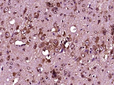
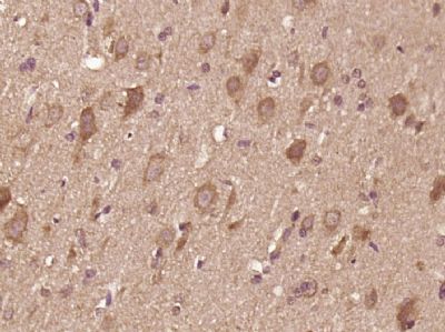
paraffin embedded (Mouse brain); Antigen retrieval by boiling in sodium citrate buffer (pH6.0) for 15min; Block endogenous peroxidase by 3% hydrogen peroxide for 20 minutes; Blocking buffer (normal goat serum) at 37°C for 30min; Antibody incubation with (IL12) Polyclonal Antibody, Unconjugated at 1:400 overnight at 4°C, followed by operating according to SP Kit(Rabbit) instructions and DAB staining.
paraffin embedded (rat brain tissue); Antigen retrieval by boiling in sodium citrate buffer (pH6.0) for 15min; Block endogenous peroxidase by 3% hydrogen peroxide for 20 minutes; Blocking buffer (normal goat serum) at 37°C for 30min; Antibody incubation with (IL12) Polyclonal Antibody, Unconjugated at 1:400 overnight at 4°C, followed by operating according to SP Kit(Rabbit) instructionsand DAB staining.
PD-L1程序性死亡配体1抗体
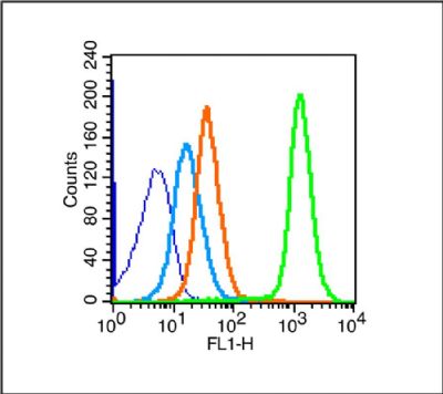
Blank control (blue line): Mouse spleen (blue).
Primary Antibody (green line): Rabbit Anti- IL12 antibody
Dilution: 1μg /10^6 cells;
Isotype Control Antibody (orange line): Rabbit IgG .
Secondary Antibody (white blue line): Goat anti-rabbit IgG-FITC
Dilution: 1μg /test.
Protocol
The cells were fixed with 70% ice-cold methanol overnight at 4℃. Cells stained with Primary Antibody for 30 min at room temperature. The cells were then incubated in 1 X PBS/2%BSA/10% goat serum to block non-specific protein-protein interactions followed by the antibody for 15 min at room temperature. The secondary antibody used for 40 min at room temperature. Acquisition of 20,000 events was performed.
Tissue/cell: rat colitis tissue; paraffin-embedded;
Antigen retrieval: citrate buffer ( 0.01M, pH 6.0 ), Boiling bathing for 15min; Block endogenous peroxidase by 3% Hydrogen peroxide for 30min; Blocking buffer (normal goat serum,at 37℃ for 20 min;
Incubation: Anti-IL-12 Polyclonal Antibody, Unconjugated 1:200, overnight at 4°C, followed by conjugation to the secondary antibody(SP-0023) and DAB staining
欢迎新老客户咨询订购:PD-L1程序性死亡配体1抗体
风险提示:丁香通仅作为第三方平台,为商家信息发布提供平台空间。用户咨询产品时请注意保护个人信息及财产安全,合理判断,谨慎选购商品,商家和用户对交易行为负责。对于医疗器械类产品,请先查证核实企业经营资质和医疗器械产品注册证情况。
 文献和实验
文献和实验曾让「白发变黑发」,Nature 研究证实这种热门的癌症疗法竟还可以抗衰老!
可能也适用于衰老细胞。 2022 年 11 月 2 日,东京大学等单位的研究人员在 Nature 发表了题为 Blocking PD-L1–PD-1 improves senescence surveillance and ageing phenotypes 的文章。该研究发现应用于癌症治疗的 PD-1 抑制剂还可以清除衰老细胞,发挥抗衰老作用。 研究表明,部分衰老细胞表达免疫检查点程序性死亡配体 1(PD-L1),PD-L1+衰老细胞在体内随年龄增长而积累。PD-L1 阴性细胞对 T
文献速递:个性化循环肿瘤 DNA 可成为免疫治疗疗效新型biomarker
研究背景 使用程序性死亡-1抑制剂或其配体(抗PD-1/PD-L1)的免疫检查点阻断 (ICB) 疗法已成为治疗许多癌症的有效方法。然而仅有少数患者对ICB疗法表现出长期良好的反应,大多数患者对免疫检查点阻断剂都没有阳性反应或因为阻断剂的不良反应而不得不停止服用。虽然已经发现了许多类别的生物标志物,但目前仍未发现强有力的治疗反应预测标志物。寻求新的ICB反应的非侵入性生物标志物能大大提高ICB疗法的临床效果。 研究内容及结果 2020年9月,在Nature子刊Nature Cancer
Nature Cancer I 生物标志物多参数评估mTLS预测实体瘤免疫治疗效果
了 TLS 中 B 细胞通过分泌抗体和细胞因子对抗 PD-1/抗 PD-L1 的反应起关键作用。DC 通过调节抗原加工和呈递来调节 T 细胞功能。B 细胞的功能依赖于 TLS 成熟度,可能是促肿瘤或抗肿瘤。在成熟 TLS 中,B 细胞和 DC 可以通过向 CD8+ 细胞提供肿瘤抗体来更好地训练 CD8+ T 细胞。和明场染色方法相比,Opal 多重免疫荧光染色同时检测更多指标,更可靠地评估 TLS 的成熟度状态。 CD3:T 细胞; CD8: 杀伤性 T 细胞;PD-L-1: 程序性死亡配体
 技术资料
技术资料暂无技术资料 索取技术资料

