

- ¥1880
- 晶风生物
- TF5551R
- 中国
- 2026年03月04日
- Elisa=1:500-1000,IHC-P=1:100-500,IHC-F=1:100-500,IF=1:100-500,ICC=1:100-500,
- Rabbit
- Human,Mouse,Rat,Dog,Pig,Cow,Rabbit,Sheep,Guinea Pig,

企业认证
相关产品推荐更多 >
万千商家帮你免费找货
0 人在求购买到急需产品
- 详细信息
- 文献和实验
- 技术资料
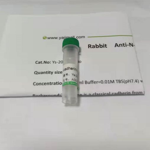
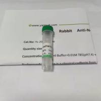
- 抗体名:
磷酸化腺苷单磷酸活化蛋白激酶α1抗体
- 抗体英文名:
phospho-AMPK α-1 (Thr183)
- 靶点:
细胞浆 细胞膜 分泌型蛋白
- 浓度:
1mg/ml
- 应用范围:
Elisa=1:500-1000,IHC-P=1:100-500,IHC-F=1:100-500,IF=1:100-500,ICC=1:100-500,
- 宿主:
Rabbit
- 适应物种:
Human,Mouse,Rat,Dog,Pig,Cow,Rabbit,Sheep,Guinea Pig,
- 保质期:
一年
- 抗原来源:
Rabbit
- 目录编号:
TF5551R
- 级别:
I级
- 库存:
10
- 供应商:
晶风生物
- 标记物:
FITC/Alexa/CY357/BIo/HRP
- 克隆性:
Polyclonal
- 保存条件:
-20
- 形态:
Liquid
- 亚型:
IgG
- 免疫原:
KLH conjugated synthetic peptide derived
- 规格:
50ul/100ul/200ul
产品名称:phospho-AMPKα-1(Thr183)磷酸化腺苷单磷酸活化蛋白激酶α1抗体
产品规格:100ul/200ul(部分有50ul,如需更大包装或其他具体规格,请咨询客服)
研究领域:肿瘤 细胞生物 免疫学等
抗体来源:Rabbit
克隆类型:Polyclonal
交叉反应:Human,Mouse,Rat,Dog,Pig,Cow,Rabbit,Sheep,
产品应用:WB=1:500-2000 ELISA=1:5000-10000 IHC-P=1:100-500 IHC-F=1:100-500 Flow-Cyt=1μg/Test IF=1:100-500 (石蜡切片需做抗原修复)
not yet tested in other applications.
optimal dilutions/concentrations should be determined by the end user.
性 状:Liquid
浓 度:1mg/ml
免 疫 原:KLH conjugated synthetic peptide derived
亚 型:IgG
纯化方法:affinity purified by Protein A
储 存 液:0.01M TBS(pH7.4) with 1% BSA, 0.03% Proclin300 and 50% Glycerol.
保存条件:Shipped at 4℃. Store at -20 °C for one year. Avoid repeated freeze/thaw cycles.
phospho-AMPKα-1(Thr183)磷酸化腺苷单磷酸活化蛋白激酶α1抗体相关抗体示例(非本抗体,如需本抗体,请联系客服索要说明书):
Sample:
Liver (Mouse) Lysate at 40 ug
Spleen (Mouse) Lysate at 40 ug
NIH/3T3 (Mouse) CellLysate at 30 ug
RAW246.7 (Mouse) CellLysate at 30 ug
Primary: Anti- IL12 at 1/300 dilution
Secondary: IRDye800CW Goat Anti-Rabbit IgG at 1/20000 dilution
Predicted band size: 22 kD
Observed band size: 35/36 kD
paraffin embedded (Mouse brain); Antigen retrieval by boiling in sodium citrate buffer (pH6.0) for 15min; Block endogenous peroxidase by 3% hydrogen peroxide for 20 minutes; Blocking buffer (normal goat serum) at 37°C for 30min; Antibody incubation with (IL12) Polyclonal Antibody, Unconjugated at 1:400 overnight at 4°C, followed by operating according to SP Kit(Rabbit) instructions and DAB staining.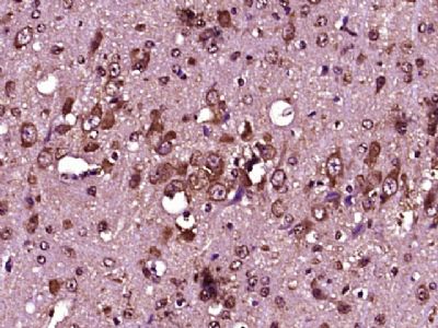
paraffin embedded (rat brain tissue); Antigen retrieval by boiling in sodium citrate buffer (pH6.0) for 15min; Block endogenous peroxidase by 3% hydrogen peroxide for 20 minutes; Blocking buffer (normal goat serum) at 37°C for 30min; Antibody incubation with (IL12) Polyclonal Antibody, Unconjugated at 1:400 overnight at 4°C, followed by operating according to SP Kit(Rabbit) instructionsand DAB staining.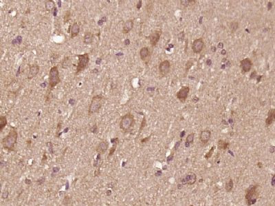
phospho-AMPKα-1(Thr183)磷酸化腺苷单磷酸活化蛋白激酶α1抗体
Blank control (blue line): Mouse spleen (blue).
Primary Antibody (green line): Rabbit Anti- IL12 antibody
Dilution: 1μg /10^6 cells;
Isotype Control Antibody (orange line): Rabbit IgG .
Secondary Antibody (white blue line): Goat anti-rabbit IgG-FITC
Dilution: 1μg /test.
Protocol
The cells were fixed with 70% ice-cold methanol overnight at 4℃. Cells stained with Primary Antibody for 30 min at room temperature. The cells were then incubated in 1 X PBS/2%BSA/10% goat serum to block non-specific protein-protein interactions followed by the antibody for 15 min at room temperature. The secondary antibody used for 40 min at room temperature. Acquisition of 20,000 events was performed.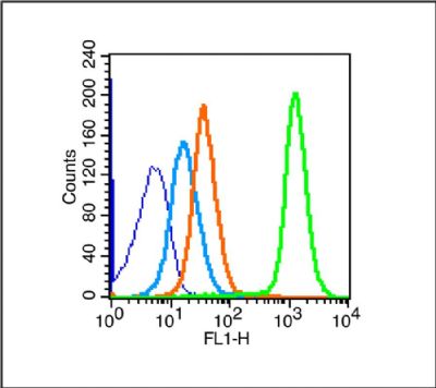
Tissue/cell: rat colitis tissue; paraffin-embedded;
Antigen retrieval: citrate buffer ( 0.01M, pH 6.0 ), Boiling bathing for 15min; Block endogenous peroxidase by 3% Hydrogen peroxide for 30min; Blocking buffer (normal goat serum,at 37℃ for 20 min;
Incubation: Anti-IL-12 Polyclonal Antibody, Unconjugated 1:200, overnight at 4°C, followed by conjugation to the secondary antibody(SP-0023) and DAB staining
欢迎新老客户咨询订购:phospho-AMPKα-1(Thr183)磷酸化腺苷单磷酸活化蛋白激酶α1抗体
风险提示:丁香通仅作为第三方平台,为商家信息发布提供平台空间。用户咨询产品时请注意保护个人信息及财产安全,合理判断,谨慎选购商品,商家和用户对交易行为负责。对于医疗器械类产品,请先查证核实企业经营资质和医疗器械产品注册证情况。
 文献和实验
文献和实验【求助】P53的翻译后修饰都有哪些? 怎样验证其修饰后与靶标启动子的结合活性的变化?
蛋白质功能的一个主要机制,p53在多个位点上可被磷酸化、顺-反异构化、乙酰化、泛素化、甲基化、糖基化等修饰,从而显示其生物学重要性。这种显示细胞或组织特异性并依赖细胞周期位置的多重修饰,是一种复杂的调节方式,随着细胞对DNA损伤、增殖、老化等产生的细胞信号的反应而发生波动。 磷酸化修饰 P53的磷酸化在多数情况下与蛋白质的稳定性有关。p53 N-末端的三个位点Ser15、Thr18、Ser20磷酸化后,使p53和其主要的负性调节因子MDM2之间的相互作用消失,而和乙
网络 第二节 蛋白酪氨酸激酶 蛋白酷氨酸激酶(protein tyrosine kinase,PTK)是一类催化ATP上γ-磷酸转移到蛋白酪氨酸残基上的激酶,能催化多种底物蛋白质酪氨酸残基磷酸化,在细胞生长、增殖、分化中具有重要作用。迄今发现的蛋白酪氨酸激酶中多数是属于致癌RNA病毒的癌基因产物,也可由脊椎动物的原癌基因产。根据PTK是否存在于细胞
第五节 蛋白激酶C 蛋白激酶C(protein kinase C,PKC)是一类Ca 2+ 、磷脂依赖性的蛋白激酶,在跨膜信号传递过程中起着重要作用。PKC通过催化多种蛋白质上Ser/Thr磷酸化,调节多种细胞的代谢、生长、增殖和分化。 表 8-3 哺乳动物组织内的PKC亚类 亚 类 氨基酸残基 分子量(kDa) 激 活 剂 表达组织 A组:经典PKC
 技术资料
技术资料暂无技术资料 索取技术资料

