大家都在搜

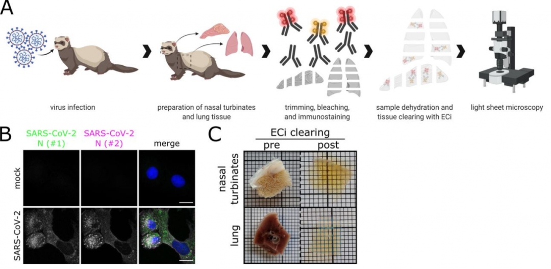
| 对于当前仍波及全球的 COVID-19,大量研究信息,如病毒的作用受体及结合位点、作用机制通过研究已经掌握;必要的研究及检测工具也在短期内得到开发;易感性研究和开发合适的动物研究模型也在多种属动物体内进行,如雪貂、仓鼠、猫、犬、貉、兔、转基因鼠、猪、猴、禽类及果蝠等。
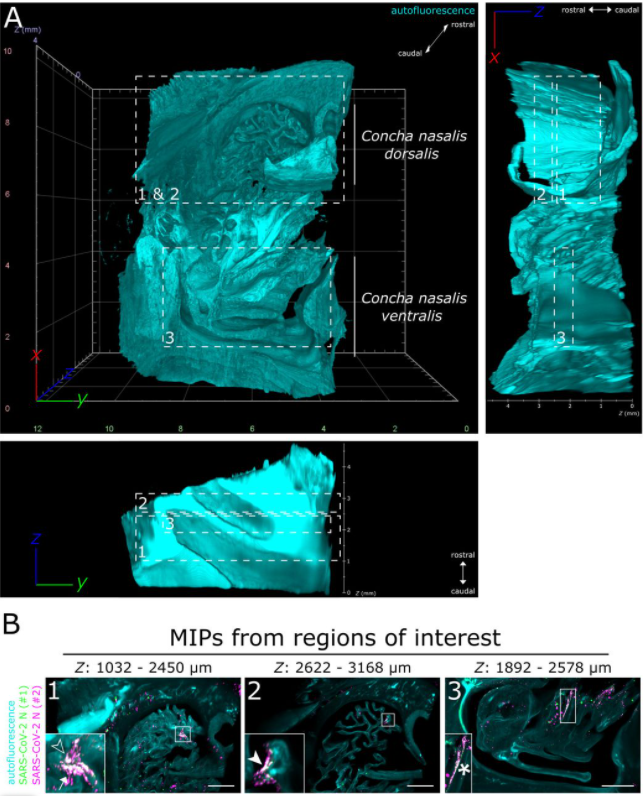
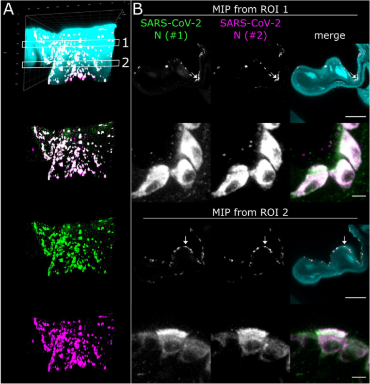
使用光片整体成像将鼻甲部位 SARS-CoV-2 感染的三维空间分布可视化。(A) 鼻甲组织结构 (> 200 mm3,感染 4 天后取材) 网格边线示意长度为 2 mm (B) 对目的区域内病毒含量进行强度投射,绿色和玫红色分别为不同来源商品化 SARS-CoV 病毒 N 蛋白。标尺 = 1 mm。 (A) 对目的区域 1(ROI 1)中 SARS-CoV-2 感染强度做投射。图像采用 40x/1.1 水浸物镜进行采集。青绿色为样本自发荧光; 绿色标记为 SARS-CoV-2(N 蛋白 1); 玫红色 = SARS-CoV-2(N 蛋白 #2)。标尺分别为 100 µm (宏观) 和 5 µm (细部)。 【1】Potratz, M.; Zaeck, L.; Christen, M.; Te Kamp, V.; Klein, A.; Nolden, T.; Freuling, C.M.; Muller, T.; Finke, S. Astrocyte Infection during Rabies Encephalitis Depends on the Virus Strain and Infection Route as Demonstrated by Novel Quantitative 3D Analysis of Cell Tropism. Cells 2020, doi:10.3390/cells9020412. 【2】Zaeck, L.; Potratz, M.; Freuling, C.M.; Muller, T.; Finke, S. High-Resolution 3D Imaging of Rabies Virus Infection in Solvent-Cleared Brain Tissue. J Vis Exp 2019, 10.3791/59402, doi:10.3791/59402. 【3】Kieffer, C.; Ladinsky, M.S.; Ninh, A.; Galimidi, R.P.; Bjorkman, P.J. Longitudinal imaging of HIV-1 spread in humanized mice with parallel 3D immunofluorescence and electron tomography. Elife 2017, 6, doi:10.7554/eLife.23282. 【4】Potratz, M.; Zaeck, L.M.; Weigel, C.; Klein, A.; Freuling, C.M.; Müller, T.; Finke, S. Neuroglia Infection by Rabies Virus after Anterograde Virus Spread in Peripheral Neurons. bioRxiv 2020, 10.1101/2020.09.20.305078, doi:10.1101/2020.09.20.305078. 【5】Ladinsky, M.S.; Khamaikawin, W.; Jung, Y.; Lin, S.; Lam, J.; An, D.S.; Bjorkman, P.J.; Kieffer, C. Mechanisms of virus dissemination in bone marrow of HIV-1-infected humanized BLT mice. Elife 2019, 8, doi:10.7554/eLife.46916. 【6】Chhatbar, C.; Detje, C.N.; Grabski, E.; Borst, K.; Spanier, J.; Ghita, L.; Elliott, D.A.; Jordao, M.J.C.; Mueller, N.; Sutton, J., et al. Type I Interferon Receptor Signaling of Neurons and Astrocytes Regulates Microglia Activation during Viral Encephalitis. Cell Rep 2018, 25, 118-129 e114, doi:10.1016/j.celrep.2018.09.003. 【7】Eckermann, M.; Frohn, J.; Reichardt, M.; Osterhoff, M.; Sprung, M.; Westermeier, F.; Tzankov, A.; Werlein, C.; Kuhnel, M.; Jonigk, D., et al. 3D virtual pathohistology of lung tissue from Covid-19 patients based on phase contrast X-ray tomography. Elife 2020, 9, doi:10.7554/eLife.60408. 【8】Li, G.; Fox, S.E.; Summa, B.; Hu, B.; Wenk, C.; Akmatbekov, A.; Harbert, J.L.; Vander Heide, R.S.; Brown, J.Q. Multiscale 3-dimensional pathology findings of COVID-19 diseased lung using high-resolution cleared tissue microscopy. |

更有优质直播、研选好物、福利活动等你来!