大家都在搜

手机验证

询价列表

暂时没有已询价产品
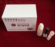
BCIP/NBT显色试剂盒(蓝
-紫)

企业认证
现货
博士德生物
Kit
| 产品货号 | AR1023 |
|---|---|
| 产品名称 | BCIP-NBT显色试剂盒/BCIP-NBT-kit |
| 价格规格 | ¥280.00/盒 |
| 试剂盒内容 | 显色剂 A:20X BCIP/NBT 浓缩液,3ml 显色剂 B:20X Tris 浓缩缓冲液,3ml |
| 产品用途 | 免疫组织(细胞)化学,原位杂交,细胞凋亡(Tunel)等实验中,碱性磷酸酶系统的显色 |
点击图片放大
风险提示:丁香通仅作为第三方平台,为商家信息发布提供平台空间。用户咨询产品时请注意保护个人信息及财产安全,合理判断,谨慎选购商品,商家和用户对交易行为负责。对于医疗器械类产品,请先查证核实企业经营资质和医疗器械产品注册证情况。